Your Image
Detector

The Apparatus

Description of image

I. The Source

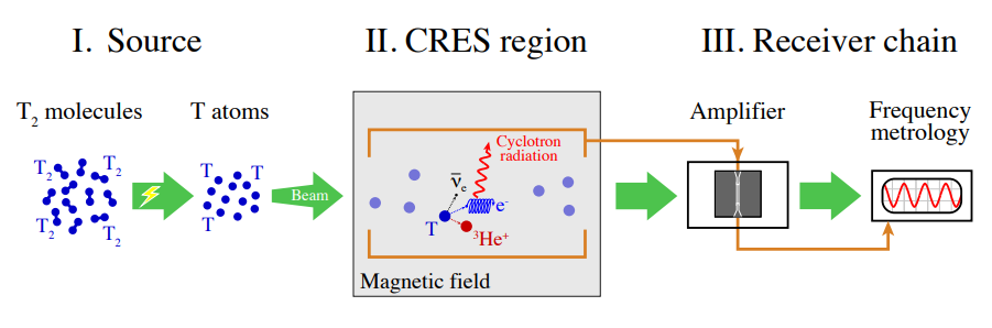
Molecular tritium introduces additional rotational and vibrational energy states, which act as a background in the energy spectrum. This background can distort the endpoint measurement, hence affecting the precision with which the neutrino mass is determined.

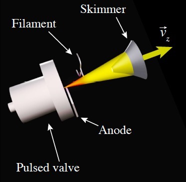
To create a simpler system for our precision measurement, we need to break down molecular tritium \(T_2\) into atomic tritium \(Tr\). This is done through a process called molecular dissociation.

Making an Atomic Tritium Source

The dissociation of molecular tritium is achieved using a DC discharge method. This process is seeded with an electron emitted from a tungsten filament. The electron initiates the dissociation process, breaking the molecular bonds and converting \(T_2\) into their atomic forms. This ensures a high-purity source of atomic tritium for the experiment.

Our experiment employs a cryogenic pulsed supersonic source operating at 30 Kelvin. This environment converts molecular tritium to atomic tritium and also minimises thermal noise, hence enhancing the signal-to-noise ratio for more accurate measurements.

The valve (pictured above) is opened, releasing a fast burst of tritium molecules. The filament, an area of high electric field, causes the tritium to dissociate/'fall apart' into atoms. These atoms possess fast forward and sideways momentum, resulting in a cone-like shape. We mainly want these to travel forward since this makes it easier for the atoms to funnel into our detector. To do this, we use a skimmer (similar to a collimator) to 'select' only those atoms travelling forward, producing a selected beam which travels through the storage ring towards the CRES region.

Tritium has a finite half-life, meaning not all tritium atoms will decay while in the detector. They continue circulating until a decay event occurs.

II. The CRES Region

Click here for more info about CRES

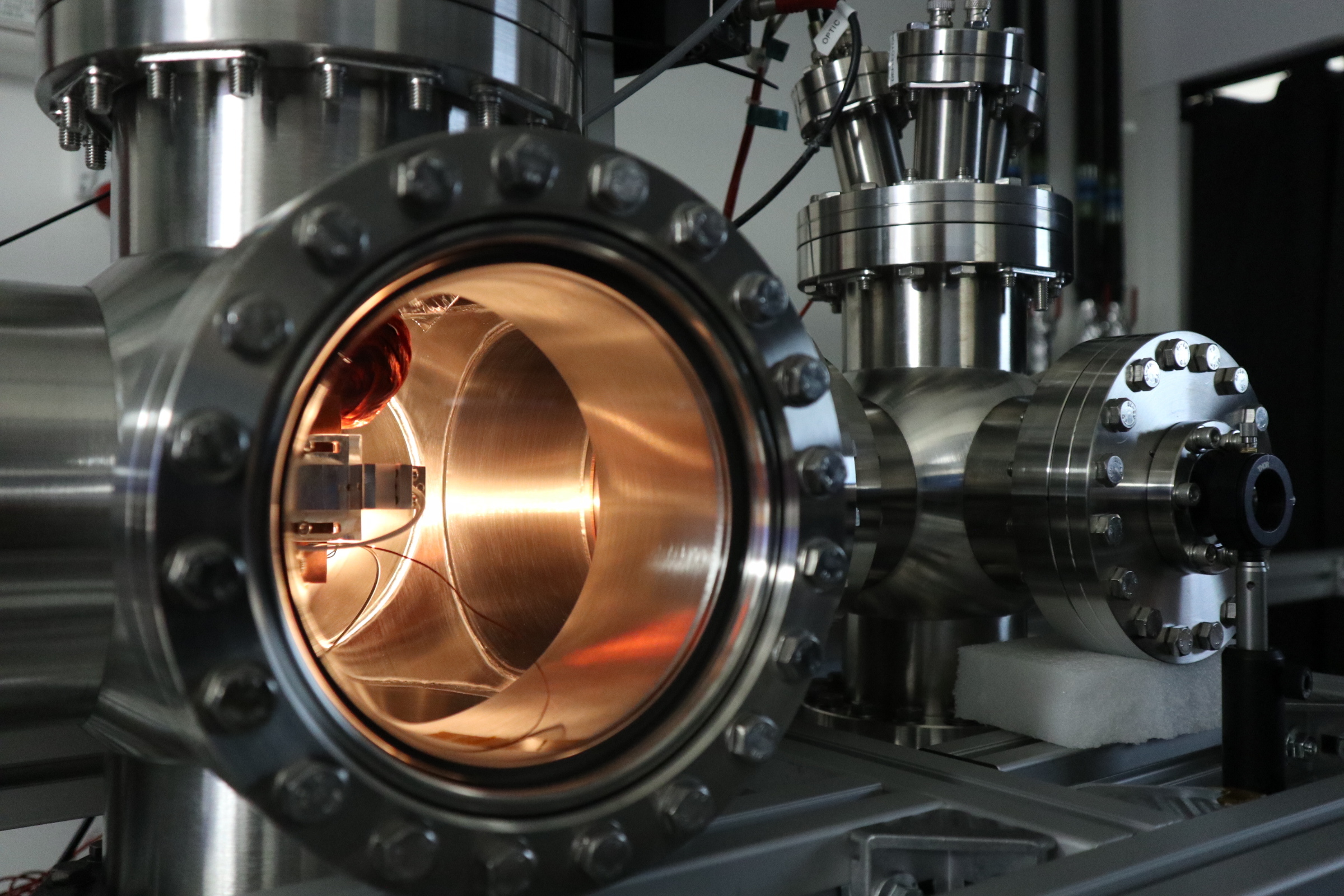
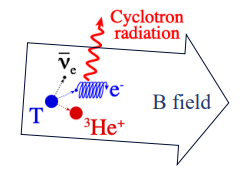
When a decay takes place, an electron is emitted into a uniform magnetic field (or B field), which is produced by a large magnet. The electron moves in a helical path within the magnetic field, emitting cyclotron radiation as it spirals.

The CRES region is where we collect this radiation.

However this comes with a challenge. The emitted electrons are incredibly fast – near-relativistic in speed – and need to be measured for >20µs in order to collect enough power to determine the kinetic energy of these electrons.

We also expect an extremely tiny signal on the order of femtowatts, with minimal thermal noise, especially since the CRES region is maintained at 4 Kelvin (almost -270°C!).

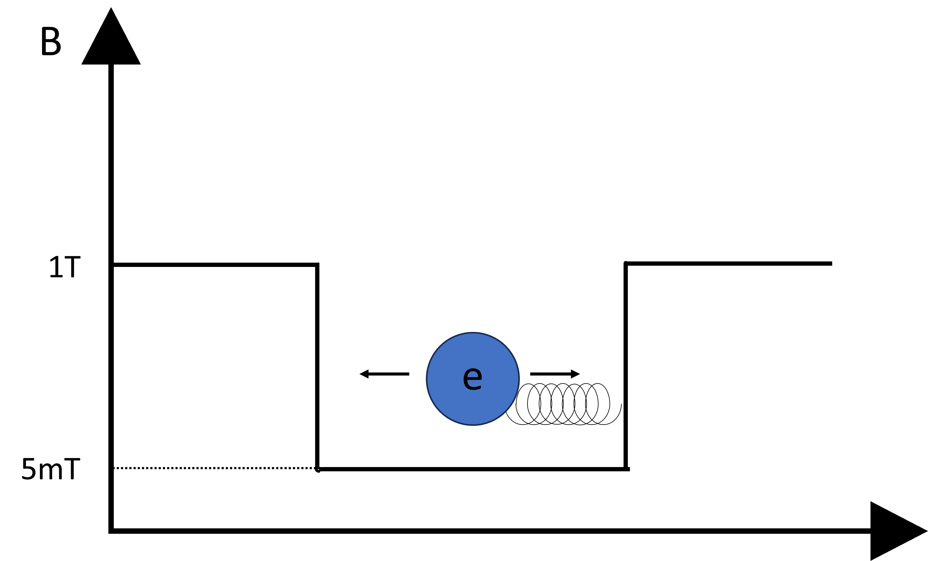
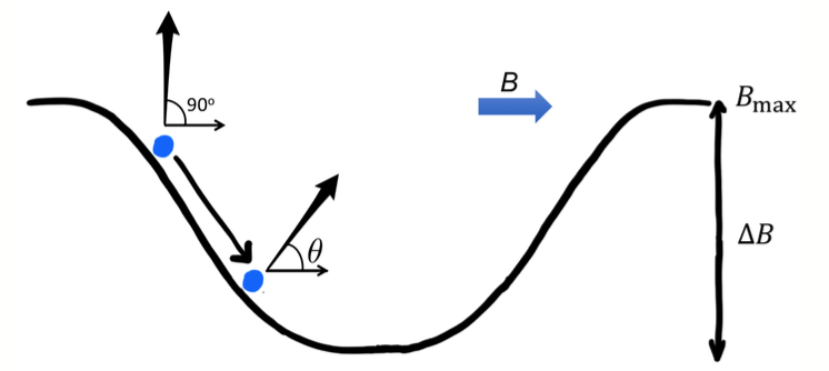
To resolve this, a 'no work' magnetic trap is placed within the uniform B field. The emitted electron bounces back and forth within this trap as it spirals, allowing us to observe it for a longer period of time.

Our "No Work Trap" Solution

Within the trap, electrons can be observed for tens or hundreds of microseconds. This 'bottle trap' design creates a local minimum in the magnitude of the background B-field.

We need a trapping field of the order of 5 mT to counteract the 1T background magnetic field.

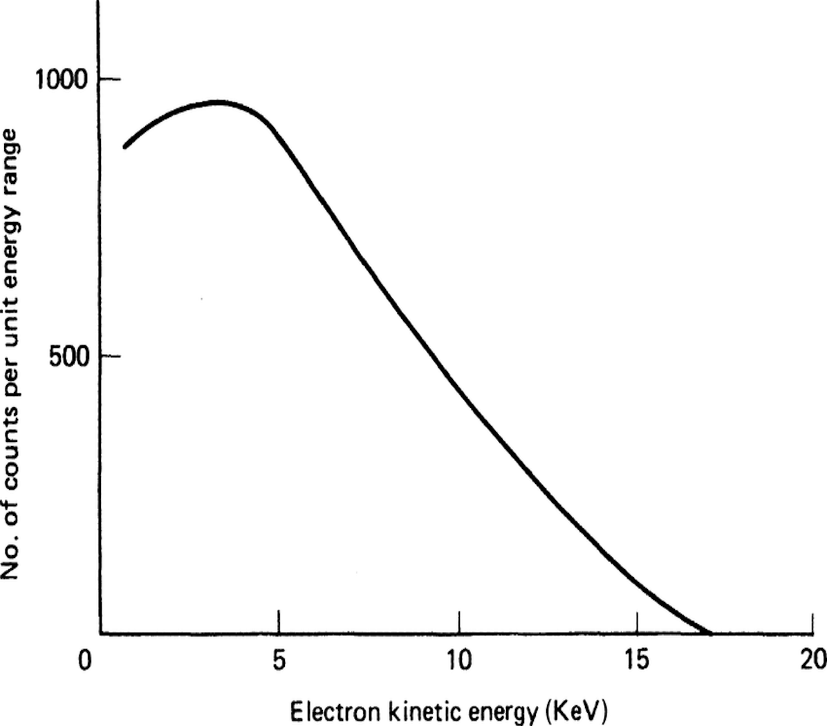
Our aim is to observe signals towards the endpoint of the spectrum - in other words, we want to make the detector 'blind' to background.

As we move towards lower energy levels, the frequency increases, meaning the chosen bandwidth (i.e. the window of frequencies observed) will affect if the signal is observed.

Radiation is collected using specialised equipment like an array of antenna or a resonant cavity.

Resonant Cavity - Since the CRES signal wavelength is ~ 1cm, we need the resonant cavity to be ~1cm long. The cyclotron radiation couples to the cavity and resonates (vibrates), giving a signal in the cavity. We can put endcaps on the cavity to adjust the bandwidth, however more cavities are required to measure more tritium. This presents a challenge since more cavities will alter the B-field since metal surfaces become charged, resulting in an irregular field.

Antenna - This method employs 'free-space detection', where the antennas would sit on the outside of the detector and 'look in', measuring the activity inside. Since we don't know where a decay will take place and hence where an emitted electron will be, an array of antennas would prove useful in detecting radiation from multiple points within the detector. These are currently being developed to 'pick up' this radiation.

Magnetometry

To precisely determine the kinetic energy of an electron in a CRES spectrometer from the frequency of its emitted radiation, it is essential to also precisely determine the strengths of the magnetic (and electric) fields in our apparatus. The measurement of such magnetic fields is known as magnetometry.

The measurement of static magnetic fields to a precision of ~\(10^{-6}\)T can be achieved in our CRES apparatus by using tritium atoms themselves, as well as other neutral atoms such as helium, as quantum sensors. In order to achieve this most effectively, these atoms must be prepared in circular Rydberg states, where the outermost electron follows a circular path around the atomic nucleus. The atoms travel through the magnetic field to be characterised at a speed of 2mm/µs (\(10^{-6}\)s)! By making measurements at a range of time delays after Rydberg state photoexcitation, the magnetic field can be mapped at a range of positions.

Pitch Angle and Average B-Field

As the tritium atoms decay, an electron will be emitted at a certain angle known as the pitch angle (θ). Electrons with a pitch angle less than 90° experience a different average magnetic field (B) due to their motion within the trap. This variation in the magnetic field can affect the frequency of emitted cyclotron radiation, thereby influencing the precision of neutrino mass measurements.

III. The Receiver Chain

The signal is so small that thermal noise is background - we want to reduce this noise (and hence increase the signal-to-noise ratio (SNR)) as much as possible.

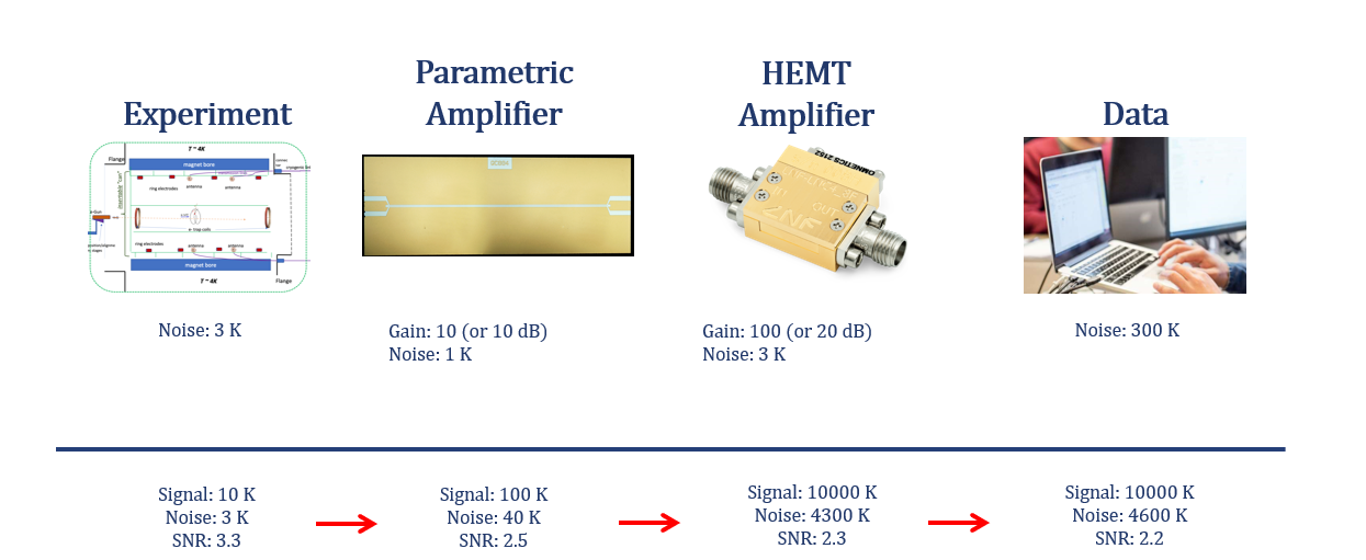
The small collected signal is amplified using a quantum amplifier, which boosts the signal power to a level where thermal noise is no longer a concern. These are also designed to operate at the fundamental noise limit, offering the potential for significant improvements in signal detection.

Quantum Limited Microwave Amplifiers

The CRES signal in the experiment is notably weak, with an approximate power of 1 femtowatt. While state-of-the-art High Electron Mobility Transistor (HEMT) amplifiers offer noise temperatures around 7K, QTNM's requirements call for the use of quantum-limited amplifiers for enhanced sensitivity. Two types of quantum-limited amplifiers are under consideration: these amplifiers operate at the quantum limit of noise performance, making it possible to detect extremely weak signals with high accuracy.

A. Superconducting Low Inductance Undulatory Galvanometers (SLUG) Amplifiers

The SLUG amplifier is a non-parametric, low-noise cryogenic amplifier currently under development at the National Physical Laboratory (NPL). Designed to operate at very low temperatures, SLUGs use niobium (Nb) nanobridge junctions to also operate at high frequencies.

Additionally, the amplifier will be fabricated from a single film of Nb, making this much simpler structurally compared to other existing methods.

B. Superconducting Parametric Amplifiers

The Cambridge Sensors Group has taken the lead in developing high-gain parametric amplifiers based on superconducting resonators, which operate at low temperatures and have lower power requirements. Superconducting Niobium Nitride (NbN) films have been chosen as the material for these amplifiers.

The current state of progress suggests that superconducting parametric amplifiers are likely to be an important component in the readout of CRES signals for the QTNM project.

QTNM will use a range of techniques from different disciplinaries disciplines, such as atomic physics, experimental physics, and quantum technology to achieve our long-term goal of determining mβ. More information about our experiment can be found in the QTNM white paper.